Alopecia Mucinosa

What is Alopecia Mucinosa?
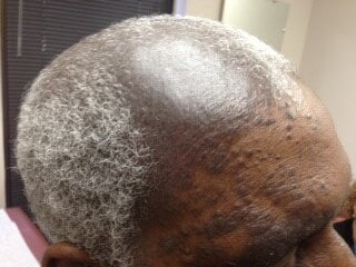
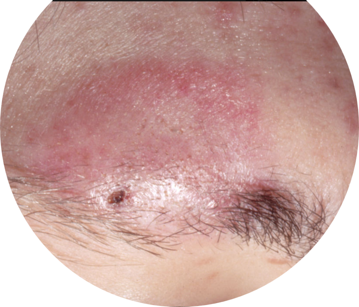
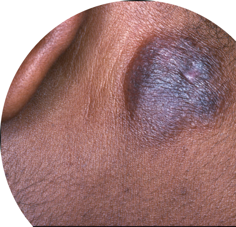
Alopecia mucinosa, also known as follicular mucinosis, is a condition characterized by the deposition of a jelly-like substance called mucin in and around the hair follicles. It can occur as a benign primary condition of unknown cause or secondary to other conditions such as atopic dermatitis (eczema) or cutaneous T-cell lymphoma. The primary form, first described in 1957 by dermatologist Pinkus, typically affects children and adults in their third and fourth decades of life, presenting as pink plaques on the face or scalp with associated scaling and hair loss. The secondary form, often a slow-growing malignancy, generally occurs in an older age group with larger areas of involvement on the extremities, trunk, and face. Diagnosis typically involves a skin biopsy, and treatment may include topical corticosteroids, intralesional corticosteroids, antimalarial therapy (hydroxychloroquine), and anti-inflammatory antibiotics such as minocycline or doxycycline.
Symptoms & Causes

Patients with alopecia mucinosa suffer from inflamed, itchy pink-red lesions over hair follicles. These lesions can present anywhere on the body but are primarily located on the head and neck region. The condition can occur with or without alopecia (hair loss) in areas with the rash; this alopecia can be scarring (permanent) or nonscarring.
There are two types of alopecia mucinosa. There is a benign, idiopathic (of unknown cause) form that arises in children and young adults with lesions centered on the head and neck that present acutely and tend to resolve spontaneously. The second type manifests more chronically in adults in their third and fourth decades of life and presents with multiple lesions spread all over the body that can be resistant to treatment. This form of alopecia mucinosa can be idiopathic but is usually secondary to a wide variety of conditions such as atopic dermatitis (eczema), lupus, and leukemias/lymphomas. The most reported association of secondary alopecia mucinosa is with mycosis fungoides, a cutaneous T-cell lymphoma.
Diagnosis & Treatments

Your dermatologist will diagnose this condition with an in-office skin biopsy. In this quick procedure, your dermatologist will administer local numbing medicine to the biopsy spot before removing a small amount of skin with a tool. They will look at this skin under a microscope to confirm the diagnosis. The overall prognosis is good, and in most cases, the condition resolves on its own within two months to two years without any treatment. In other cases, patients may require treatment to prevent future hair loss and manage symptoms like itching, scaling or redness.
There is no one specific treatment option. Topical, intralesional (injected directly into the lesion), and systemic corticosteroids can be helpful. Additional therapeutic methods include isotretinoin, antimalarial therapy (hydroxychloroquine), antibiotics (minocycline, doxycycline, or dapsone) and UVA1 light therapy. In cases secondary to an underlying disease or malignancy, therapy should be focused on treating that condition. Overall, different therapies will work for different patients, and it is recommended for individuals to follow up consistently for at least five years after diagnosis in case of malignancy.
