Folliculitis Decalvans

What is Folliculitis Decalvans?
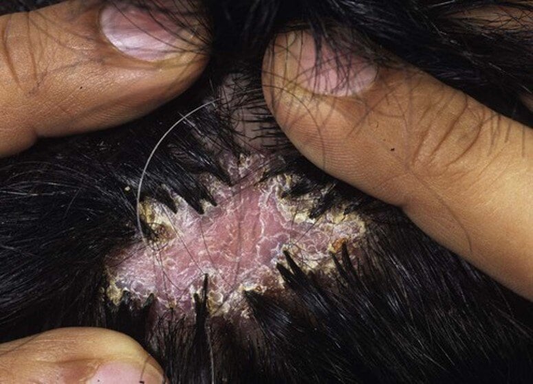
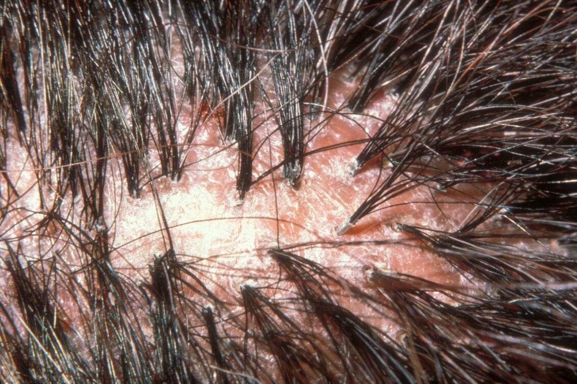
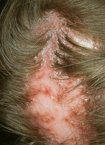
Folliculitis decalvans belongs to a group of disorders called cicatricial or scarring alopecias that destroy the hair follicle, replace it with scar tissue, and eventually causes permanent hair loss. Decalvans is a term derived from the Latin meaning “making bald.” Folliculitis decalvans is further classified as a neutrophilic scarring alopecia, based upon the type of inflammation (neutrophils) seen on routine biopsies in the early stages of the disease. This condition typically starts with crops of red bumps around the base of the hair follicles, which then develop marked inflammation, pustules formation, crusting and eventually patchy hair loss.
Symptoms & Causes

Cause
The cause of folliculitis decalvans is unknown, however it has been speculated that the disease reflects an abnormal host response to bacteria, most commonly Staphylococcus aureus. Such organisms may act as “superantigens” and may escape detection by the host immune system. Interestingly, patients with folliculitis decalvans typically do not have evidence of bacterial infection elsewhere on the skin, nor do they have any evidence of immune deficiency.
Signs & Symptoms
Although extensive epidemiologic studies are not available, the incidence of folliculitis decalvans comprises about 11% of all scarring alopecia cases. Folliculitis decalvans typically occurs in young and middle-aged adults with a slight male predominance. It also occurs more frequently in African Americans compared to Caucasians. Folliculitis decalvans can be seen families, thereby supporting a genetic predisposition. The initial lesions of folliculitis decalvans involves a red bump around the base hair follicle usually along the crown or posterior scalp. As the inflammatory process continues bright inflammation, pus formation, scaling, and crusting takes place. Patients occasionally report spontaneous bleeding and frequently complain about pain, itching, and/or burning sensations. As the disease progresses small discrete to extensive, irregularly shaped smooth scarred patches of hair loss develop.
Diagnosis & Treatments

Experienced dermatologists will suspect the diagnosis of folliculitis decalvans from a detailed clinical history, thorough physical examination of the scalp, bacterial cultures, and a scalp biopsy. It is important to know if the patient has a history of recurrent S. aureus or other bacterial infections. A scalp biopsy the gold standard of diagnosis of scarring hair loss and are often performed when considering a diagnosis of folliculitis decalvans.
The most helpful information from the biopsy is the type and pattern of inflammation (especially predominance of neutrophils) and the presence of bacterial pathogens. A bacterial culture from a pustule or a tissue culture from the scalp tissue can be helpful in determining the best treatment plan. Early diagnosis and therapeutic intervention can often prevent permanent damage to hair follicles. Once a definitive diagnosis is made, we can determine the best treatment plan for everyone. Some regrowth is even possible with treatment.
Since bacteria (most commonly Staphylococcus aureus) seems to play an important role in the pathogenesis of folliculitis decalvans, appropriate and long-term antimicrobial therapy directed at the predominant pathogen is needed to control the disease process. Repeated culture of pustules is needed to guide the choice of antimicrobial agents, and this may change over time. Several different oral antibiotics, such as the tetracyclines, trimpethoprim-sulfamethoxale, and erythromycin have been shown to be beneficial. Rifampin 300 mg twice daily over 10–12 weeks is believed to be the best treatment for S. aureus eradication and may lead to successful long-term remission. It is strongly recommended to use rifampin in combination with clindamycin 300 mg twice daily to avoid rapid emergence of resistance. Topical antibiotics such as mupirocin, clindamycin or topical anti-septics such as chlorhexidine can be helpful in conjunction with oral antibiotics. Topical corticosteroids can help decrease inflammation and calm associated symptoms (itching, redness, or pain). Topical tacrolimus may also be helpful as a steroid sparing topical agent. Additionally, injections of corticosteroid, such as triamcinolone acetonide, may be used in inflamed and symptomatic areas, every 4-6 weeks. Other oral anti-inflammatory agents that can be used include oral corticosteroids (for short periods of time), oral isotretinoin, and dapsone.
