Frontal Fibrosing Alopecia

What is Frontal Fibrosing Alopecia?
Frontal Fibrosing Alopecia (FFA) is an inflammatory skin disorder with progressive hair loss (alopecia) due to the destruction of hair follicles. The hair loss is centered around the eyebrows and front hairline and can be permanent or scarring in nature. FFA was first discovered in 1994 and is classified as a subtype of a condition called lichen planopilaris. In the past decade, incidence rates of FFA have been increasing globally. While this condition can occur in anyone, it most commonly presents in peri and postmenopausal women.
Symptoms & Causes

The cause of frontal fibrosing alopecia is still unclear. It is believed to be a complex inflammatory process likely induced by both genetic and environmental factors. Recently, researchers discovered that the disease has been linked to a gene associated with the formation of estrogen, which suggests why women undergoing hormonal shifts surrounding menopause are most affected. There have also been some reports of FFA occurring within families, further suggesting a genetic cause. Research also suggests that FFA can be triggered by cosmetic products (sunscreen, makeup, facial soap), sun exposure, and psychological stress. However, there has been no proven cause-and-effect relationships between environment factors and frontal fibrosing alopecia. Therefore, more research is needed on this topic. Additionally, studies have shown the disease is associated with many other autoimmune diseases, such as hypothyroidism.
Like its parent diagnosis, lichen planopilaris, the target of inflammation is the sebaceous or oil gland which is located at the same site as the stem cell region of the hair follicle. Recent research has shown that there is a defect in a “master regulator” protein called the peroxisome proliferator-activated receptor gamma, or PPAR gamma. PPAR gamma is responsible for the preservation of hair follicle cells, including stem cells, and sebaceous glands. Loss of PPAR gamma leads to sebaceous gland dysfunction, which causes abnormal processing and buildup of “toxic” lipids. This abnormal buildup of lipids triggers inflammation that destroys the oil gland inflammation and the nearby stem cells, which are innocent bystanders. Once stem cells are destroyed, the follicles cannot regenerate, and permanent hair loss ensues.
The typical profile of a person with frontal fibrosing alopecia is a peri or post-menopausal adult female, with an average age of 56-63 years. It can also occur in premenopausal women, even as young as teenagers, as well as adult men, with an average age of 47 in this population.
Because of slow progression, the disease often goes unnoticed for many years before it is diagnosed. There is a triad of findings in fibrosing alopecia which includes:
(1) Sudden eyebrow loss. This can be diffuse or patchy loss, and most of the time is accompanied by redness and scaling around the hair follicles. The existing eyebrow hairs themselves may change texture and become course and wiry. Overtime complete permanent eyebrow loss can occur.
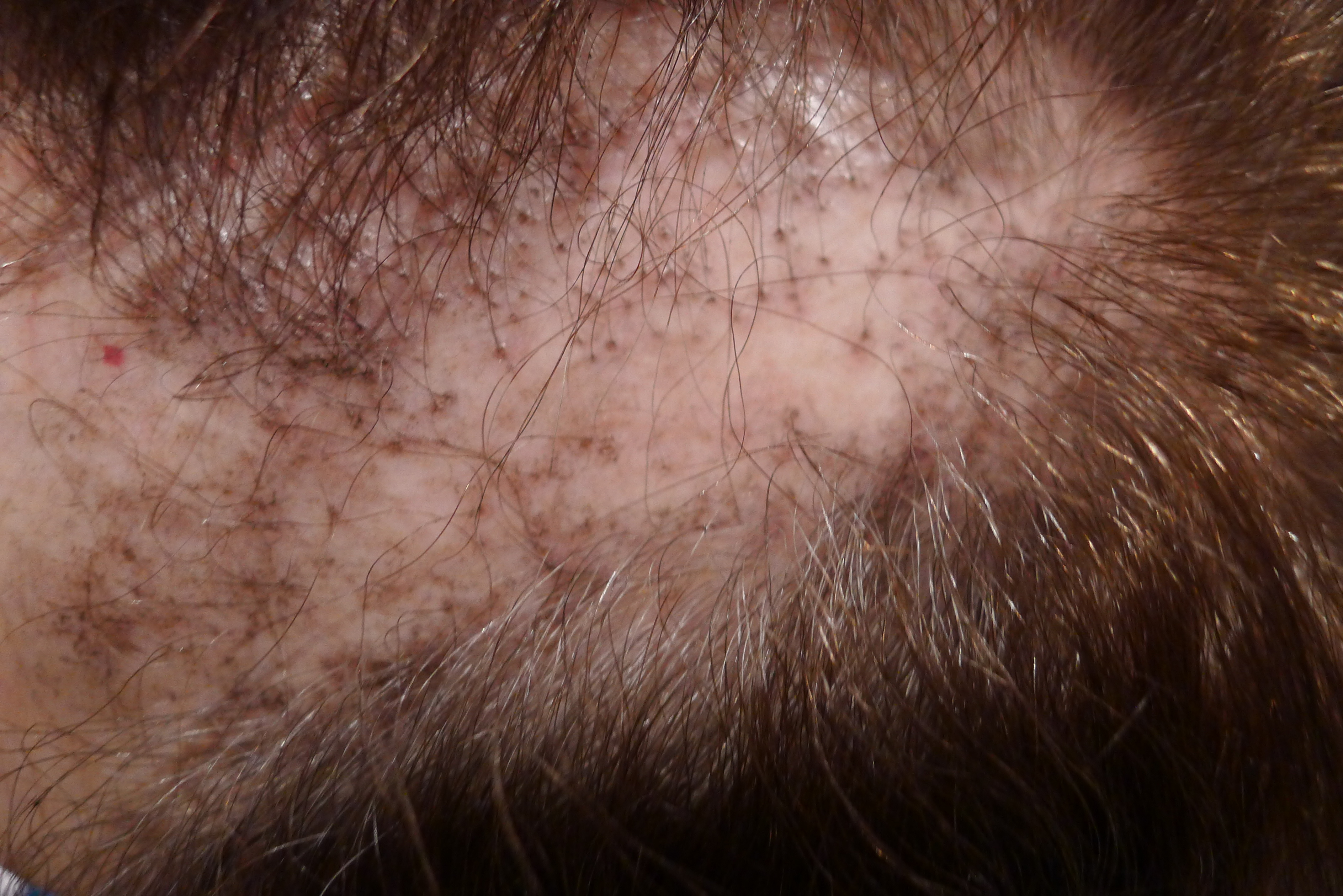
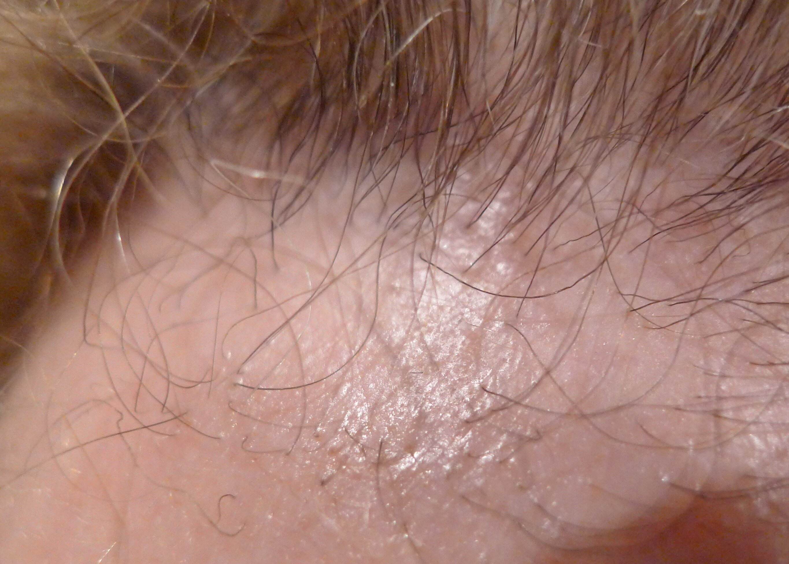
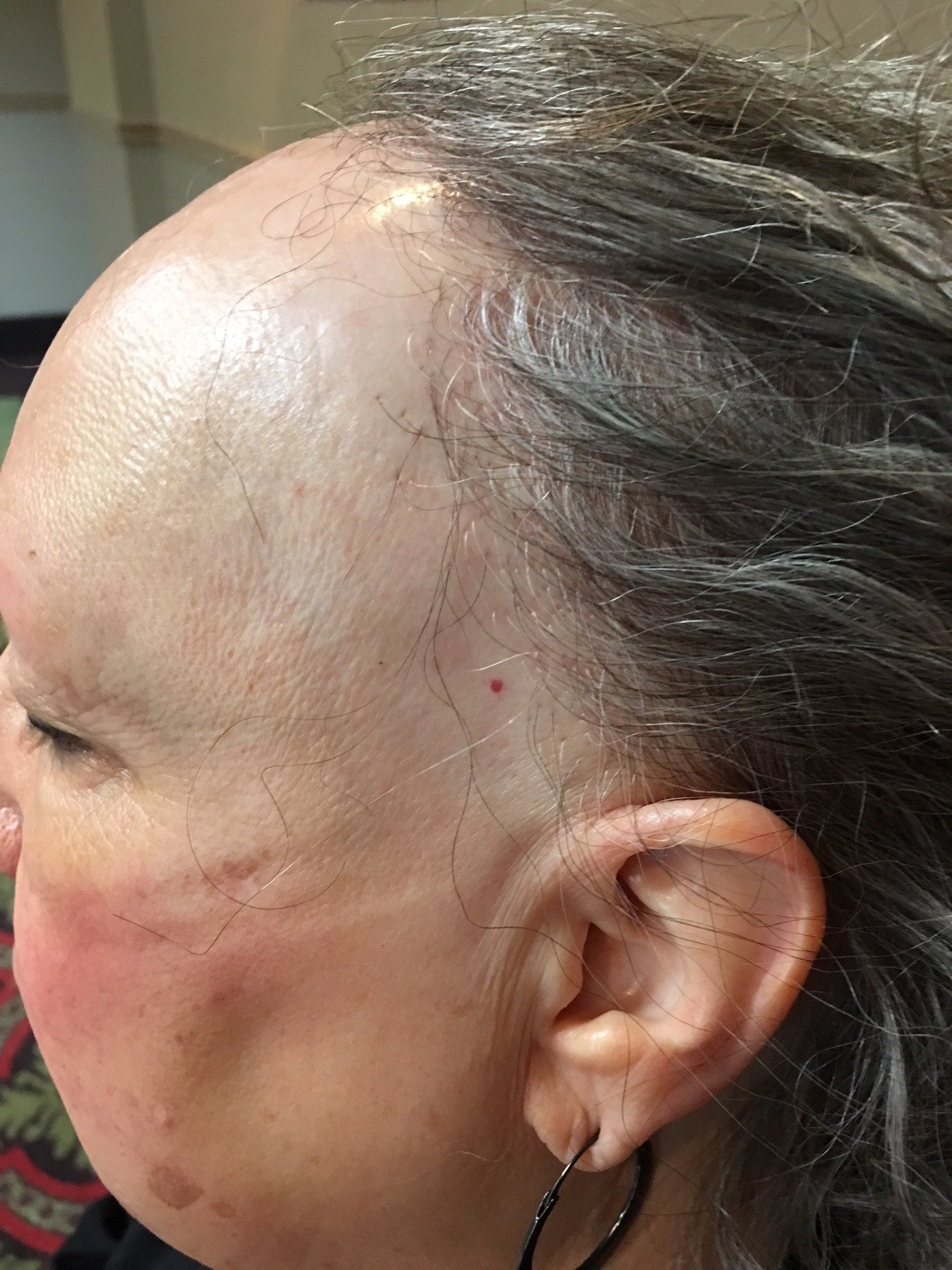
(2) Hairline recession. As the name of the disease implies, the frontal hairline is most often affected, followed by the sideburns, above and around the ears and even the posterior hairline. The hair loss spans across the front of the head in a “band-like” pattern like a receding hairline. The hair follicles surrounding the hair loss have characteristic redness and scaling.
(3) Extremity hair loss. This is a more inconspicuous finding because extremity hair loss can happen naturally with age. However, hair loss on the extremities, like arms and legs, in frontal fibrosing alopecia can be patchy and completely bald which is unlike age related hair loss.
There are three typical patterns of hair loss in frontal fibrosing alopecia:
1) Pattern I (linear)- a linear and uniform receding hairline, without any loss of hair behind the new hairline. This can present with eyebrow loss.
2) Pattern II (diffuse)- diffuse or zigzag alopecia all across the frontal hairline with significant loss of hair behind the new hairline as well. This also can present with eyebrow loss.
3) Pattern III (pseudo-fringe)- a band of alopecia behind the original frontal hairline, called the “pseudo fringe sign.” Notably, eyebrows remain unaffected.
Patients may present with atypical hair loss that does not fall in one of those three patterns. Other findings of frontal fibrosing alopecia include skin-colored facial bumps, or papules. Sometimes frontal fibrosing alopecia can overlap with classic lichen planopilaris.
Diagnosis & Treatments

Your dermatologist will diagnose this condition with an in-office skin biopsy. In this quick procedure, your dermatologist will administer local numbing medicine to the biopsy spot before removing a small amount of skin with a tool. They will look at this skin under a microscope to confirm the diagnosis. Scalp biopsies are critical for the assessment and diagnosis of scarring hair loss. Early diagnosis and therapeutic intervention can prevent permanent damage to existing hair follicles. Once a definitive diagnosis is made, the best treatment plan can be determined. Some regrowth is even possible with treatment.
Management of frontal fibrosing alopecia is complex as there is no consensus on treatment guidelines. Treatment is case-dependent, based on severity of symptoms (itching and inflammation), findings on biopsy and extent of hair loss. Response to treatment varies from individual to individual. In many cases, the disease spontaneously stabilizes without further hair loss.
In mild disease, topical potent corticosteroid solutions may halt the progression of hair loss. However, in most cases, topical treatments alone are not sufficient. Intralesional corticosteroids, which are steroids injected directly into the spot of hair loss, have been shown to be effective in promoting hair regrowth. This treatment option may not be optimal if hair loss is more diffuse, as the steroids only work in the spot they were injected, or due to the adverse risk of wearing down skin (atrophy).
For more diffuse or severe cases, 5-alpha-reducatase inhibitors like finasteride (Propecia) have shown to be the most effective in treatment. The anti-diabetic drug pioglitazone (Actos) is also a promising treatment option due to its stimulating effects on PPAR gamma. Previous studies suggest oral anti-inflammatory medications like doxycycline and hydroxychloroquine (Plaquenil) could be beneficial; however, the evidence is not strong. In some cases, immunosuppressant medications such as azathioprine (Imuran), mycophenolate mofetil (Cellcept) and cyclosporine can be used to achieve control. Lastly, patients with stabilized disease may qualify as good candidates for hair graft transplants but should be followed up for at least three years post graft due to risk of relapse.
